FORAMENSTENOSE
Ungeklärte Lahmheit der Hintergliedmaßen bei Tieren
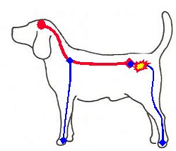
Der Krankheitskomplex Foramenstenose, einige sprechen auch von Nervenwurzelkompressionssyndrom, betrifft die Hintergliedmaßen des Tiers und löst Rückenschmerzen sowie bewegungsabhängige Nervenwurzelschmerzen aus, die strangförmig durch den Oberschenkel über die Wade bis hinunter in die Pfote ziehen. Beschreibungen dieses Schmerzmusters sind nicht nur bei Tieren, sondern auch vom Menschen bekannt, er kann ebenfalls an Foramenstenose leiden.
Betroffene Tiere werden meist mit einem Vorbericht monatelang anstehender Lahmheiten bei mehreren Tierärzten vorstellig. Die orthopädischen Voruntersuchungen sind in der Regel umfassend und gut, jedoch ohne Diagnose. Die gängigen Schmerzmittel (NSAID) sind meist unwirksam, Kortison hilft nur vorübergehend.
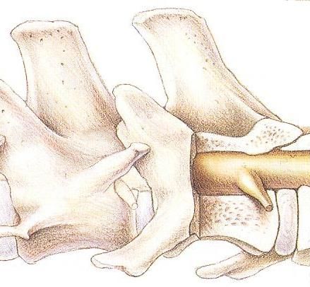
Anatomie
Bei Menschen wie bei Tieren verlassen die Nervenwurzeln des Rückenmarks durch Neuroforamen (kleine Aussparungen zwischen Wirbelkörpern) die Wirbelsäule, um sich außerhalb der schützenden Wirbelsäule zu Nerven zusammenzufinden, die dann in der Peripherie gelegene Körperregionen versorgen.
Auf Druck oder Zug reagieren diese Nervenwurzeln sehr schmerzempfindlich (Nervenwurzelschmerz).
Erscheinungsbild der Krankheit
Tiere mit Foramenstenose sind neurologische Patienten, ihr Nervenwurzelschmerz stellt sich jedoch so dar, als habe der Hund oder die Katze ein orthopädisches Problem (Bewegungsschmerz ohne Lähmung).
Oft lassen die Tiere ein kurzes Aufjaulen oder eine Schmerzäußerung im Sprung nach oben hören, auch kann ein Anheben der Gliedmaße schmerzhaft sein, da der Ischiasnerv dabei gestreckt wird. Arbeit verschlimmert, Ruhe verbessert den Schmerz. Eine Streckung im Übergang der Lendenwirbelsäule zum Kreuzbein (wie dies bei einem Sprung nach oben geschieht) verstärkt Schmerzen und/oder die Lahmheit.
Die Tiere stehen nicht so fest auf dem krankem Bein, fortgeschrittene Fälle zeigen eine verminderte Muskelspannung und sogar Muskelschwund (beim Abtasten Gefühl der „leeren Hose“). Patienten mit beidseitiger Foramenstenose stellen sich ständig von einem Bein aufs andere. 20% der Patienten haben unabhängig von der Foramenstenose zusätzlich Hüft- und Knieprobleme.
Inkontinenz und Harnabsatzstörungen können in Verbindung mit der Foramenstenose auftreten und auch nach Behandlung derselben wieder verschwinden.
In unserer Tierarzt-Praxis beobachten wir eine Häufung bei 2-11-jährigen Jagdhunden bzw. Hunden der Rassen Deutscher Schäferhund, Labrador Retriever, Boxer, Dogge, Hovawarth, Briard, Border Collie.
Diagnostik der Foramenstenose
Da sich ein Neuroforamen im Röntgenbild unzureichend, der Nerv gar nicht darstellt, entgehen diese Patienten lange ihrer Diagnose.
Die Nervenwurzel wird selten so stark eingeengt, dass ihre motorischen Funktionen betroffen sind. Daher sieht man selten Lähmungserscheinungen an der betroffenen Gliedmaße.
Für eine saubere Diagnose untersuchen wir das Tier sehr sorgfältig klinisch und neurologisch. Dabei wenden wir Röntgen- und Elektrodiagnostik sowie Kernspintomographie an. Dies ist auch im Hinblick auf eine exakte Operationsplanung von herausragender Bedeutung. Begleitende Erkrankungen des Tieres müssen zuvor erkannt werden!
Es kommt nicht zu einer Spontanheilung der Foramenstenose! Ohne Operation schreitet der Prozess sicher weiter voran!
Operation (Foraminotomie)
In der tierärztlichen Operation wird das Neuroforamen so geweitet, dass die Nervenwurzel wieder druck- und verklebungsfrei durch die Wirbelsäule verlaufen kann. Anschließend benötigen die Tiere Physiotherapie und innerhalb von 3-6 Monaten wird bei ca. 80% der Patienten eine Beschwerdefreiheit erreicht (Bereiche, in denen die Nervenwurzel druckbedingt ihre Nervenscheide verloren hat, werden langsam wieder in funktionelle Strukturen regeneriert).
Physiotherapie bei Tieren nach Foraminotomie
Einen beachtlichen Teil des Behandlungsergebnisses verdanken wir einer effizienten Physiotherapie. Der Tier-Physiotherapeut behandelt verspannte, schmerzhafte Muskeln, die operationsbedingt entstehen oder durch Fehlhaltung (Gewichtsverlagerung weg vom Schmerz nach vorn) krankheitsbedingt schon vor dem Eingriff vorhanden waren.
Diese Programme beinhalten:
1.-2. Woche passives Training
3.-6. Woche Muskeltraining, Leinenzwang
6.-12.Woche Traben, Wassertraining, Terraintraining
4.-5. Monat Konditionstraining
Arbeitshunde bekommen den Hürdensprung erst nach (4)-6 Monaten nach gutem Aufwärmen erlaubt.[:en]