ELECTRODIAGNOSTICS
Our electrodiagnostics unit provides us with an extremely useful tool for carrying out nerve and muscle function tests. Hearing tests (audiometry) and eye tests are also available.
How does such a device work?
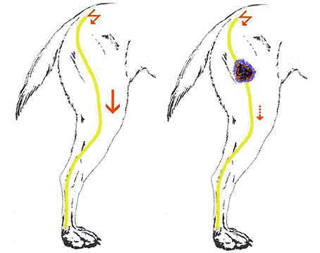
The electrodiagnostic unit measures the finest electrical signals and discharges that control the body. When a stimulus originates in the brain, for example, it travels along the spinal cord via the nerves to a muscle, which then moves when the stimulus reaches it. With the help of electrodiagnostics, such stimuli can also be generated artificially and then it is possible to measure how much of this stimulus reaches the muscle and how long it takes for such a stimulus to reach it. Delays due to nerve damage can thus be easily detected.
Stimulation activities in the muscles can also be measured and interpreted in order to draw conclusions about nerve and muscle diseases.
For practical use, this is a great help in recognizing and classifying neurological problems and thus helps to better diagnose neurological patients and document healing successes.
Often, however, it is also unclear lameness, the causes of which can be clarified by using electrodiagnostics. Some patients find it difficult to distinguish between orthopaedic and neurological gait disorders on the basis of the clinical examination. How electrodiagnostics can make a decisive contribution to confirming the diagnosis is described below (“Nerve and muscle function tests”).
The chapters on “Hearing tests” and “Vision tests” explain how an animal’s vision and hearing can be determined and documented, even on one side.
Nerve and muscle function tests
Diseases of tendons, joints or bones cause gait disorders that are classified as orthopaedic. This is referred to as lameness.
A disease of the nerves or muscles leads to a neurological gait disorder. This is referred to as paralysis. In some patients, lameness and paralysis cannot be distinguished with the naked eye and on clinical examination. However, as the treatment of both can differ considerably, differentiation with the aid of electrodiagnostics is important.
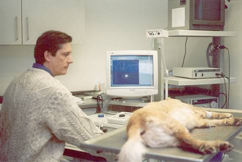
 Here electromyography (EMG) and a Animal neurology: electrodiagnostic examination of a dog by Dr. Florian König help to make an unequivocal diagnosis. Neurological patients show spontaneous activity in the musculature 5-7 days after the damage has occurred. The muscles, which are undersupplied with nerve signals, begin to send out signals independently, which can be measured with EMG. Such a phenomenon is not observed in orthopaedic lameness.
 Furthermore, the ability of each major nerve to transmit electrical impulses can be measured. It is also possible to measure the speed at which a nerve transmits electrical impulses. This type of nerve function analysis not only helps to identify nerve damage, but also to localize it and assess its severity. A basic prerequisite for a targeted prognosis and therapy!
Hearing tests (Brain Auditory Evoked Potentials, BAEP, audiometry)
Deafness is not only inherited in Dalmatians. This is why we often carry out breeding tests, which give breeders the certainty that they are handing over hearing-healthy puppies to their new owners. However, the question of hearing ability also arises in many other patients.
The ability to hear can be impaired by damage to the ear or auditory nerve (peripheral) or by damage to the brain (central), which processes the stimuli, converts them and allows them to become conscious. The distinction between these two forms is of great importance for the prognosis, i.e. the assessment of whether the animal can learn to hear again or not.
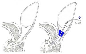
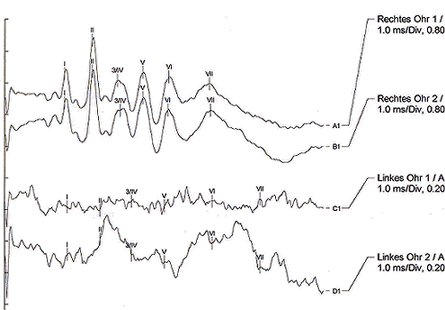
When measuring BAEP, acoustic signals (clicks) are sent into the ear to be examined and the reaction to these signals (spiral ganglia, auditory nerve, brain stem) is recorded on the scalp (similar to brain wave measurements). This works from the age of 5 weeks. The BAEP can be used to localize, make an unequivocal statement and document it. In Dalmatians and many other breeds, this is a prerequisite for breeding approval; in many other animals, the BAEP provides clarity and diagnostic certainty.

Schematische Darstellung eines Hörtests

Hörtest einer linksseitig tauben Katze
Vision tests (visual evoked potentials, VEP, electroretinography, ERG)
When assessing vision, three structures are of interest: the eye, which receives the visual stimulus, the optic nerve, which transmits it, and the brain, which processes it and makes it conscious. Damage to one of these three units can lead to the same clinical manifestation (blindness). Since different diseases are treated differently, we naturally want to know where the blindness is triggered.
We use VEP to measure brain waves that indicate a reaction in the brain after a light stimulus has been administered. If these stimuli are processed in the brain, we can assume that there are functioning pathways from the eye via the optic nerve (peripheral) to the brain (central). This not only means that the animal can receive the visual stimuli via the eye, but can also transmit them and then process them in the brain. An interruption of these functions is indicated by the VEP.
The ERG measures the absorption of light stimuli by the eye. This is essential information when it comes to deciding, for example, whether lens surgery (cataracts) is advisable in order to restore vision. This is because only an otherwise intact eye is able to see again after lens surgery.
In many cases, foregoing new diagnostic services is no longer appropriate and often undesirable.
Author: Dr. Florian König
